Retinal Vein Occlusion Q&A
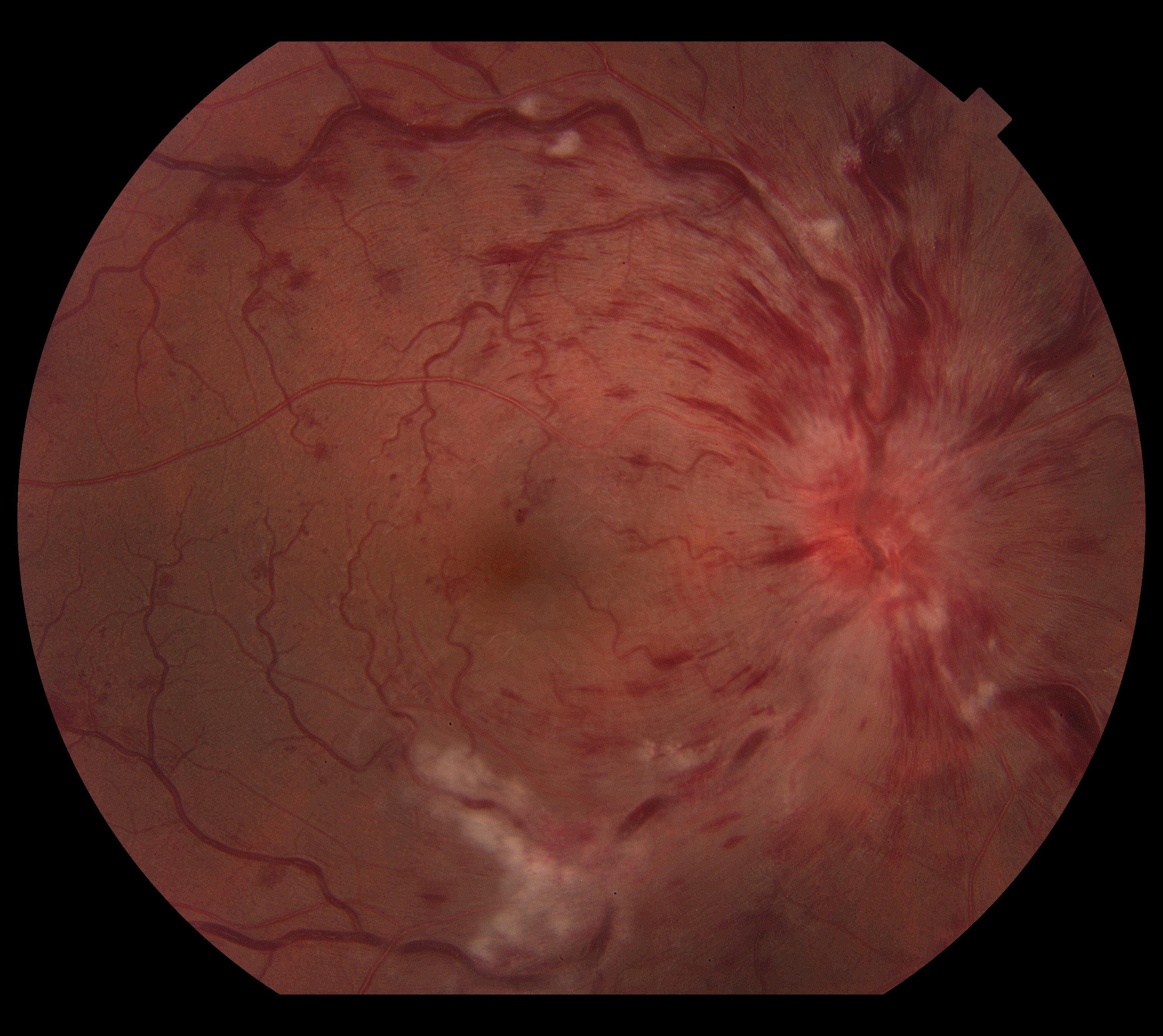
- Central Retinal Vein Occlusion (CRVO)
- Branch Retinal Vein Occlusion (BRVO)
Clear, practical answers to common retinal vein occlusion questions.
Jump to a question
Tap a question to jump straight to the answer.
- What is a retinal vein occlusion?
- What causes a retinal vein occlusion?
- What symptoms should I watch for?
- How do I use an Amsler grid?
- Will my vision recover?
- How is it diagnosed?
- What is macular oedema and why is it important?
- How is retinal vein occlusion treated?
- Will I need injections? How often?
- Will I just need one treatment of injection for macular oedema from a retinal vein occlusion?
- How frequently will i need my eye injections?
- Will I ever be able to stop my injections for macular oedema from a retinal vein occlusion?
- Are the injections painful or risky?
- What are the risks of an eye injection?
- What should I do after my injection?
- Can this happen in my other eye?
- Do I need medical tests after this diagnosis?
- Can I still drive?
- Is this condition permanent?
- What should I do if my vision suddenly worsens?
Q
What is a retinal vein occlusion?
A retinal vein occlusion occurs when one of the veins draining blood from the retina becomes blocked. This causes blood and fluid to leak into the retina, which can affect vision. If the main vein is blocked, it is called a Central Retinal Vein Occlusion (CRVO). If a smaller branch vein is blocked, it is called a Branch Retinal Vein Occlusion (BRVO).
Q
What causes a retinal vein occlusion?
It is usually caused by a blood clot forming in a retinal vein. Vascular risk factors include high blood pressure, diabetes, high cholesterol, smoking, and glaucoma. It is more common as we get older. Managing these conditions reduces risk.
Q
What symptoms should I watch for?
Symptoms often include sudden blurred or reduced vision in one eye. Some people notice dark spots or distortion. CRVO tends to cause more severe vision loss than BRVO. Sudden changes should always be checked urgently. An Amsler grid is a simple tool you can use at home to help monitor your central vision and detect early changes. If you notice any new distortion or missing areas on the grid, contact your eye doctor promptly.
Q
How do I use an Amsler grid?
To use an Amsler grid, wear your reading glasses (if you use them) and hold the grid at your normal reading distance in good light. Cover one eye and focus on the central dot with the uncovered eye. While staring at the dot, notice whether any of the surrounding lines look wavy, blurred, distorted, or missing. Repeat the test with the other eye. If you see new distortion, dark patches, or missing areas, contact your eye doctor promptly. Try to check your vision with the grid regularly, such as once a week.
Q
Will my vision recover?
Recovery depends on how severe the blockage is. Some patients, especially with BRVO, experience partial improvement. Others may have permanent vision changes. Early treatment improves the chances of better outcomes.
Q
How is it diagnosed?
It is diagnosed during a dilated eye examination. Special retinal scans (OCT) and sometimes dye tests are used to assess swelling and blood flow. These tests are painless and help guide treatment.
Q
What is macular oedema and why is it important?
Macular oedema is swelling in the central retina caused by fluid leakage. It is the most common cause of vision loss in vein occlusions. Treating the swelling is key to improving or stabilising vision.
Q
How is retinal vein occlusion treated?
Treatment usually focuses on reducing macular swelling and optimizing your underlying vascular risk factors. Treatment of the macular oedema often, but not always, involves anti-VEGF injections into the eye. In some cases, steroid injections or laser treatment may be used. Treatment depends on the severity. You will also need to see your GP for blood pressure checks, blood tests, and cardiovascular risk assessment. Controlling general health reduces future risk to a recurrence in the affected eye or in the other eye.
Q
Will I need injections? How often?
If you have macular oedema from a retinal vein occlusion, you may need regular injections, especially at the beginning of treatment. These may start monthly and then reduce in frequency depending on your response. The schedule is tailored to your eye’s condition. Ongoing monitoring is important.
Q
Will I just need one treatment of injection for macular oedema from a retinal vein occlusion?
No, treatment for macular oedema is usually not a one-off injection. Most patients require a series of injections, especially at the beginning, to reduce the swelling in the macula. Treatment often starts with monthly injections and is then adjusted depending on how your eye responds. Some people may need ongoing injections long term to keep the fluid under control, while others may be able to space treatments further apart. Regular monitoring is essential, as the macular oedema can recur if treatment is stopped too early.
Q
How frequently will i need my eye injections?
The frequency of your eye injections depends on how your macula responds to treatment. Most patients begin with 4-weekly injections until the fluid in the macula has settled and the condition is stable. Once the macula swelling improves, we gradually extend the interval in 2-week steps—for example, from 4-weekly to 6-weekly, then 8-weekly—provided there is no sign of recurrence. Every patient is given a careful trial of extension, but not everyone can tolerate longer gaps between injections. The longest interval we usually extend to is 16 weeks, as long as the eye remains stable. Regular monitoring is essential to keep your vision protected.
Q
Will I ever be able to stop my injections for macular oedema from a retinal vein occlusion?
Yes, it is possible to stop injections for macular oedema in some patients. This depends on how well your eye responds to treatment. If the swelling in the macula settles and your diabetes is well managed, injections may be reduced or even stopped. Good diabetes control of your vascular risk factors plays a major role in reducing the need for ongoing treatment.
Q
Are the injections painful or risky?
The eye is numbed before treatment, so discomfort is minimal. Most patients tolerate injections very well. Serious complications are rare. Your doctor will explain the risks.
Q
What are the risks of an eye injection?
Eye injections (such as anti-VEGF injections) are commonly performed and generally very safe, but like any procedure, they carry some risks. The most serious—though rare—risk is infection inside the eye (endophthalmitis), which can threaten vision and requires urgent treatment. Other uncommon risks include bleeding, retinal detachment, inflammation, increased eye pressure, or cataract formation. More common and mild side effects include temporary redness, irritation, watery eyes, or the feeling of something in the eye for a day or two. Your doctor takes careful precautions to minimise these risks, and serious complications are uncommon.
Q
What should I do after my injection?
After your injection, your eye may feel slightly irritated or watery for a day or two. It is very important not to rub your eye, as this can increase the risk of infection. You should also avoid swimming, hot tubs, or getting pool or ocean water in your eye for at least one week after the injection. Continue using any prescribed drops as directed, and monitor for symptoms such as increasing pain, worsening vision, or significant redness—if these occur, contact your doctor immediately.
Q
Can this happen in my other eye?
It is possible, especially if underlying risk factors are not controlled. Managing blood pressure, diabetes, cholesterol, and avoiding smoking reduces risk. Regular eye checks are important.
Q
Do I need medical tests after this diagnosis?
Yes. Because retinal vein occlusion is linked to vascular health, your doctor may recommend seeing your GP for blood pressure checks, blood tests, and cardiovascular risk assessment. Controlling general health reduces future risk.
Q
Can I still drive?
Many patients can continue driving if vision in at least one eye meets legal standards. Severe vision loss may affect driving ability. Your doctor can advise you based on your test results.
Q
Is this condition permanent?
The blockage itself cannot be reversed, but the complications (like swelling) can often be treated. Some vision loss may be permanent. Early treatment and good medical care give the best outcomes.
Q
What should I do if my vision suddenly worsens?
If you notice a sudden drop in vision, increased floaters, pain, or new distortion, contact your eye doctor immediately. Prompt assessment is important to protect your vision.